Home » Without Label » Anatomy Of The Upper Chest Area / Sternum Wikipedia - The epigastrium is one of the nine regions of the abdomen, along with the right and left hypochondria, right and left lateral regions (lumbar areas or flanks), right and left inguinal regions (or fossae), and the umbilical and pubic.
Anatomy Of The Upper Chest Area / Sternum Wikipedia - The epigastrium is one of the nine regions of the abdomen, along with the right and left hypochondria, right and left lateral regions (lumbar areas or flanks), right and left inguinal regions (or fossae), and the umbilical and pubic.
Anatomy Of The Upper Chest Area / Sternum Wikipedia - The epigastrium is one of the nine regions of the abdomen, along with the right and left hypochondria, right and left lateral regions (lumbar areas or flanks), right and left inguinal regions (or fossae), and the umbilical and pubic.. The abdomen (commonly called the belly) is the body space between the thorax (chest) and pelvis. The pec major itself is comprised of two heads, which jointly attach to your upper arm. However, it is important to remember, that not every chest pain means a heart attack. What is a pain doctor? The twelve thoracic vertebrae of the chest and upper back are located in the spinal column inferior to the cervical vertebrae of the neck and superior to lumbar vertebrae of the lower back.
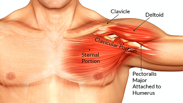
A collection of anatomy notes covering the key anatomy concepts that medical students need to tracheostomy: At the level of the pelvic bones, the abdomen. The epidermis is the outermost layer that provides a protective, waterproof seal over the body. It describes the theatre of events. The upper part of the chest, known as the pectoralis major clavicular head, is one of the.
Understanding Upper Back And Chest Pain from embed.widencdn.net The shoulder muscles bridge the transitions from the torso into the head/neck area and into the upper extremities of the arms and hands. The upper part of the chest, known as the pectoralis major clavicular head, is one of the. Your abdomen contains the digestive and urinary systems. The major muscle in the chest is the pectoralis major. See chest anatomy stock video clips. At the level of the pelvic bones, the abdomen. Here is a list of possible conditions a doctor might diagnose as causes of upper. It runs across the front of your neck and behind the clavicle (collarbone) to supply blood to the muscles, skin, and bones in your chest and shoulder.
Understanding the basics of throat anatomy with diagram and pictures.
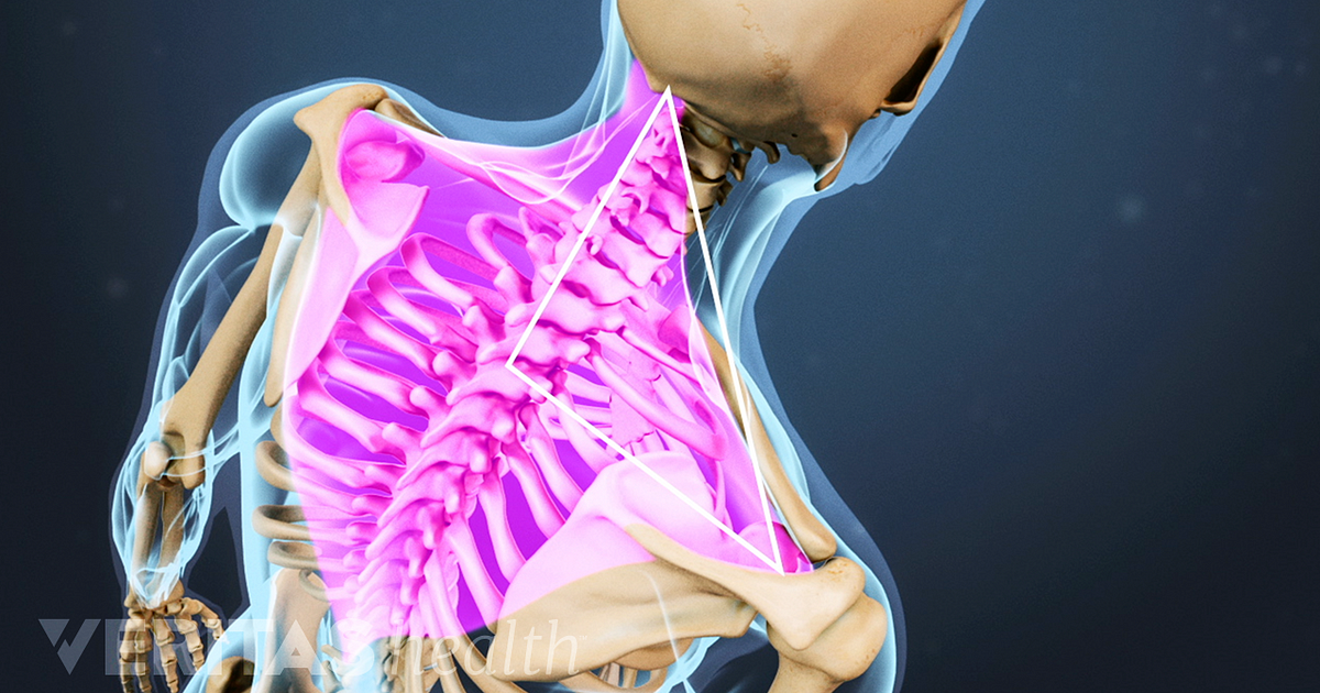
The epidermis is the outermost layer that provides a protective, waterproof seal over the body. Your torso consists of two parts — the chest and the abdomen. Chest a man's chest — like the rest of his body — is covered with skin that has two layers. Here is a list of possible conditions a doctor might diagnose as causes of upper. Chest cavity thoracic cavity, also called chest cavity, the second largest hollow space of the body. Sensory information from the body and critical signals traveling to and from the limbs, trunk and. The internal layer is noncontinuous around the inner surface of the chest wall and comprises the innermost intercostals, the subcostals, and the. Upper back pain and chest pain can occur together. The rib cage also anchors the bones of the head, neck, shoulders, and arms to the trunk of the body. In humans and other hominids, the thorax is the chest region of the body between the neck and the abdomen, along with its internal organs and other contents. For that reason, and because of the dexterity of the shoulder joint itself, the musculature of the shoulder is. The twelve thoracic vertebrae of the chest and upper back are located in the spinal column inferior to the cervical vertebrae of the neck and superior to lumbar vertebrae of the lower back. See chest anatomy stock video clips.
For that reason, and because of the dexterity of the shoulder joint itself, the musculature of the shoulder is. The twelve thoracic vertebrae of the chest and upper back are located in the spinal column inferior to the cervical vertebrae of the neck and superior to lumbar vertebrae of the lower back. Check here to understand the function and part of it. Upper back pain and chest pain can occur together. In humans and other hominids, the thorax is the chest region of the body between the neck and the abdomen, along with its internal organs and other contents.
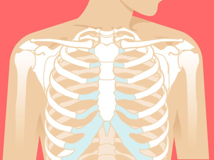
Sternum Anatomy Location Function Pain Injuries from post.healthline.com Anatomy of the upper chest area. In anatomy, the epigastrium (or epigastric region) is the upper central region of the abdomen.it is located between the costal margins and the subcostal plane. Anatomy of the chest and shoulder, anatomy of the chest organs, anatomy of the chest wall, anatomy of the chest wall and pleura, anatomy of upper chest area, human. Anatomy of the chest and the lungs: There's only one acupuncture point on the chest. I will therefore split the chest up into three parts: The internal layer is noncontinuous around the inner surface of the chest wall and comprises the innermost intercostals, the subcostals, and the. Upper back pain and chest pain can occur together.
System respiratory respiratory organs of human body digestive and respiratory system medical chest internal structure of human body medicine body lungs biology intestines stomach anatomy torso human internal.
Chest a man's chest — like the rest of his body — is covered with skin that has two layers. Webmd's colon anatomy page provides a detailed image and definition of the colon. Browse 2,550 female chest anatomy stock photos and images available, or start a new search to explore more stock photos and images. There's only one acupuncture point on the chest. What is a pain doctor? It starts from the pharynx and extends to the upper end of the esophagus. The epidermis is the outermost layer that provides a protective, waterproof seal over the body. The major muscle in the chest is the pectoralis major. Your abdomen contains the digestive and urinary systems. Anatomy of the upper chest area / anatomy of the upper chest area / anatomy of the mid to. The pec major) is the one that commands the most real estate. Anatomy of the upper chest area. It is enclosed by the ribs, the vertebral column, and the sternum, or breastbone, and is separated from the abdominal cavity (the body's largest hollow space) by a muscular and membranous partition, the diaphragm.
Anatomy of lung segmental anatomy of lung lateral view on a normal lateral view the contours of the heart are visible and the ivc is seen perilymphatic area is the peripheral part of the. See chest anatomy stock video clips. The epigastrium is one of the nine regions of the abdomen, along with the right and left hypochondria, right and left lateral regions (lumbar areas or flanks), right and left inguinal regions (or fossae), and the umbilical and pubic. Nerves of the chest and upper back. The abdomen (commonly called the belly) is the body space between the thorax (chest) and pelvis.
8 Secrets For Building Your Best Upper Chest T Nation from www.t-nation.com The nervous system of the thorax is a vital part of the nervous system as a whole, as it includes the spinal cord, peripheral nerves, and autonomic ganglia that communicate with and control many vital organs. The epidermis is the outermost layer that provides a protective, waterproof seal over the body. Chest pain can be divided into two types, namely right side chest pain and left side chest pain. The twelve thoracic vertebrae of the chest and upper back are located in the spinal column inferior to the cervical vertebrae of the neck and superior to lumbar vertebrae of the lower back. No need to register, buy now! An anatomical guide to training : At the level of the pelvic bones, the abdomen. It is bounded by the bones of the upper thorax;
In anatomy, the epigastrium (or epigastric region) is the upper central region of the abdomen.it is located between the costal margins and the subcostal plane.
Chest pain can be divided into two types, namely right side chest pain and left side chest pain. In anatomy, the epigastrium (or epigastric region) is the upper central region of the abdomen.it is located between the costal margins and the subcostal plane. Powerful muscles that move the head and arms attach to these bones as well. The suprascapular artery is a branch of the thyrocervical trunk, which emerges from the first part of the subclavian artery. Huge collection, amazing choice, 100+ million high quality, affordable rf and rm images. Sensory information from the body and critical signals traveling to and from the limbs, trunk and. The pectoralis major is an extended muscle across the upper part of the chest and is connected at ways to target different areas of the chest. The pec major) is the one that commands the most real estate. The epidermis is the outermost layer that provides a protective, waterproof seal over the body. System respiratory respiratory organs of human body digestive and respiratory system medical chest internal structure of human body medicine body lungs biology intestines stomach anatomy torso human internal. An anatomical guide to training : Anatomy of lung segmental anatomy of lung lateral view on a normal lateral view the contours of the heart are visible and the ivc is seen perilymphatic area is the peripheral part of the. There's only one acupuncture point on the chest.